.svg?sfvrsn=be606e78_3)
In order to remove a polyp or adenoma, you first have to see it. New technologies ranging from low-tech mechanical innovations to high-tech digital solutions enhance the views of specific areas of the colon and reduce the number of adenomas that remain hidden from view behind folds and flexures. Increasing adenoma detection rates (ADR) requires visualizing all of the colon’s mucosa (mucosal exposure) and being able to better spot adenomas on the mucosa you see (highlighting). That calls for a combination of higher video resolution, wider viewing angles and — let’s not forget this — good technique.
- Home
- Article
Boost Your Adenoma Detection Rates
By: Joe Paone
Published: 4/3/2019
Share:
The latest scope upgrades enhance views of the colon to highlight hard-to-spot growths.
1. Better image quality
If you’re still using standard-def scopes, you’re likely behind the curve in ADR. High-definition scopes are a must now, due to their ability to much more clearly display small polyps. It’s far easier for physicians to detect adenomas thanks to the added detail these vastly improved cameras provide — 5 times the pixel count of standard definition. Analyses show that HD gives you a 3% to 4% gain in ADR, according to gastroenterologist Douglas K. Rex, MD, a professor at Indiana University-Purdue University in Indianapolis, Ind. This enhanced view can be particularly helpful for less experienced examiners. A key point, though: Just having an HD scope won’t help unless your whole system is HD-enabled, including the processor and the video monitor.
We’re starting to see some ultra-high-def 4K scope technology hit the market, and that even further-enhanced image quality — 4 times the resolution of HD — certainly can’t hurt. But as with HD, all elements of the video processing chain must be 4K-capable to derive its benefits.
2. Mechanical scope attachments
Colonoscopes are long, thin and flexible, and not specifically designed to look behind folds and flexures in the colon wall. That’s led to some inventions that enhance the basic scope design. Mechanical extensions can help scopes explore hard-to-see areas of the colon. Combining these mucosal exposure devices with an HD scope system can help improve ADR.
- Cap-assisted. This is as rudimentary as it gets. You’re placing a transparent plastic cap on the distal tip of the colonoscope. The idea is that the cap will flatten mucosal folds.
- Cuffs and rings. Other options are more elaborate than simple caps, but are likewise attached to the distal end of colonoscopes. One is a hard plastic device featuring a single row of hinged, flexible arms. The other is designed with layers of flexible silicone-rubber rings. The devices’ flexible arms and rings fall flat against the scope during intubation and expand during withdrawal to flatten folds, stabilize the scope and prevent the scope from slipping while the physician examines the colon’s mucosa.
- Balloon-assisted. Reusable balloon attachments can be connected to standard scopes and inflated with a foot pedal once the scope has reached the cecum. When the scope is withdrawn, the balloons steady the scope and flatten folds in the colon.
These attachments are intended to improve ADR, and studies have found they do to varying degrees. However, your facility’s results might vary. For example, Ned Snyder, MD, MACP, AGAF, chief of gastroenterology at the Kelsey-Seybold Clinic in Houston, Texas, says, “We have intermittently used the [cuff] on the end of the scope, [but] it has been difficult to demonstrate benefit.”
3. Image enhancements
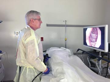
CLEARLY BETTER High-definition imaging is just one of the advancements that have improved the effectiveness of colonoscopy screenings.
Beyond physical attachments, you can also improve your views of the colon with high-tech approaches that highlight adenomas.
- Virtual/electronic chromoendoscopy. Chromoendoscopy, which uses dye stains to highlight abnormalities in mucosa, has never really caught on outside of inflammatory bowel disease because of the hassle associated with preparing it, notes Dr. Rex. But the concept obviously has merit as a highlighting technology for colonoscopy.
Virtual chromoendoscopy, also known as electronic chromoendoscopy, is not new to colonoscopy. It was largely ineffective in its original form due to lighting issues, but newer versions, which have brighter illumination, make the technique more effective, says Dr. Rex. There are competing technologies available — narrow-band imaging and blue light imaging being the most prominent.
The idea here, again, is to provide better contrast than traditional white light would; chromoendoscopy lets you better spot adenomas because they show up as a different color than the mucosa around them. But instead of using chemical dyes, you’re achieving a similar effect by filtering wavelengths of light. Adenomas emit different wavelengths than the mucosa around them, and these devices can differentiate and display them.
- Wide-angle colonoscopes. The concept here is worthy. As opposed to a forward-viewing colonoscope with a 140- to 170-degree viewing angle, these multi-camera devices approach full 360-degree views. Through either an add-on to a standard scope or with a fully-equipped specially designed scope, a pair of side-facing cameras complement the forward-viewing camera. The resulting images are combined to provide a panoramic view of the mucosa.
When evaluating wide-angle scopes, however, keep in mind that they might not have the same resolution quality as conventional HD scopes.
- Artificial intelligence. Based on machine learning, AI has great potential as a highlighting technology to improve detection. Leveraging data consisting of tens of thousands of mucosal images, an AI-enabled scope would predict the locations of potential lesions and draw a box around those mucosal areas so that the physician can take a closer look.
AI will probably be available with the next generation of scopes, says Dr. Rex. But will it be an add-on, or will there be special AI scopes? Also, he says questions remain about its actual sensitivity and specificity, or how often it will produce false positives. “Bottom line, we don’t have enough data right now to know how good [AI is] going to be in the next couple years,” says Dr. Rex. “Will the technology eventually be good enough to make it standard? Absolutely. But we still have to see results from clinical trials before that happens.”
Expanding the scope
CMS is increasingly using ADR as a reference for evaluating and compensating gastroenterologists. According to CMS, physicians should have an ADR of at least 20% in women and 30% in men. While you can justifiably debate the fairness and accuracy of the metric — given the variables in patient age, susceptibility and other factors that individual doctors experience in their unique practices — it’s the new reality. But it’s also true that, from a patient care perspective, the more polyps your physicians find, the better.
Many of these newer technologies designed to improve ADR can also improve both cecal insertion and withdrawal times, and that could positively impact your efficiencies. If you save 2 or 3 minutes per exam, and you’re doing 15 exams a day, you might be able to schedule an additional screening, get some other work done or end your day a bit earlier, says Dr. Rex.
There are also less tangible benefits to optimizing colono-scopy screenings. “From a societal standpoint, increasing ADR helps decrease the cost of cancer care,” adds Dr. Rex. “And for individual examiners, it’s the peace of mind of having performed a better examination.” OSM
.svg?sfvrsn=56b2f850_5)