December 17, 2020

Share:
eNews Briefs December 17, 2020

THIS WEEK'S ARTICLES

4 Tips for Preventing Wrong-Site Spine Surgery

Spine Surgeon Has Close Encounter with Shark

Advanced Practice Practitioner Took Lead to Streamline Care of Patients With Painful Spine Fractures

When Spine Outcomes Don't Match Patient Perception

A New ERAS in Spine Surgery Care


4 Tips for Preventing Wrong-Site Spine Surgery

They range from effective verbal communication to the latest navigation systems.

INCLUSIVE CULTURE 
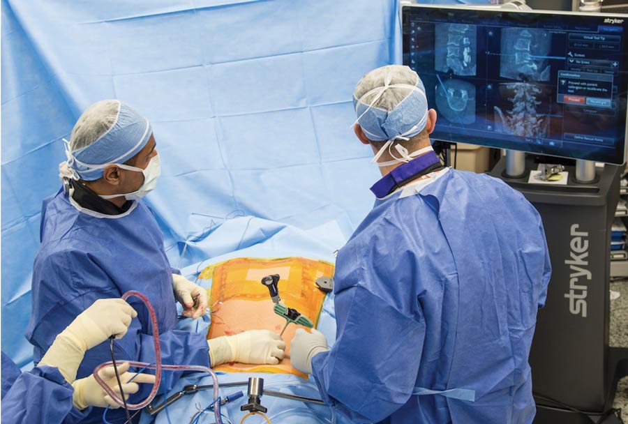
Credit: Hospital for Special Surgery
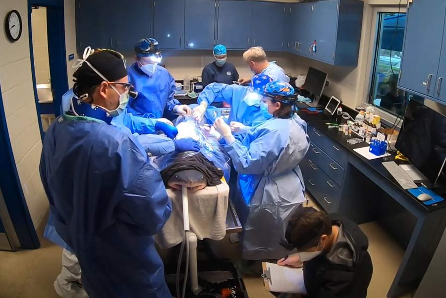
INCLUSIVE CULTURE When surgeons count vertebrae, OR staff should feel empowered to speak out if they see a possible error.

In the delicate world of spine surgery, a wrong-site error can be disastrous both for the patient and your facility. The causes are varied, including similar-looking vertebrae, patient obesity, anatomic abnormalities and visualization limitations. Sheeraz Qureshi, MD, MBA, the Patty and Jay Baker endowed chair in minimally invasive spine surgery at the Hospital for Special Surgery, and associate professor of orthopedic surgery at Weill Cornell Medical College in New York City, shares four essential tips that help surgeons more accurately identify the level of the spine that needs to be repaired or replaced:

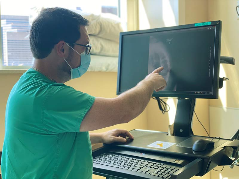
  • Make three counts. Because individual vertebrae can look identical, identifying the correct level of the spine to be operated on can be difficult. "The only way to know you're at the correct spot is to find an anatomical landmark that looks different from the rest of the spine, and count vertebrae until you reach the disc that needs treatment," says Dr. Qureshi. C-arms don't provide full-body images, so he places artificial markers to identify where to begin his count to the correct vertebrae. He performs three localized counts using X-rays to confirm the intended surgical site. First, he does a pre-incision count after the patient is intubated, anesthetized and positioned; he places a needle through the skin that overlays the area he wants to operate on and confirms it's at the vertebrae that needs repair. Second, he performs a post-incision count by placing a surgical tool on the bone to make sure it's the correct one; his count ends when he gets to the disc with the tool on top. A third count for confirmation is taken after the surgery.
  • Involve the whole OR team. Dr. Qureshi advises to conduct time outs pre-incision, post-exposure and post-surgery. "Every member of the surgical team should be engaged in ensuring the correct site is identified," he says. "They should stop what they're doing when it's announced a localization attempt is about to start and focus all of their attention on the patient, the surgeon and the intraoperative images." He counts out loud to the targeted vertebrae level, and if someone doesn't agree, he encourages staff to question him and ask for another image or count if necessary. Dr. Qureshi recommends all OR staff receive advanced training about counts, localizations and spinal anatomy. "All it takes is a culture change," he says. "Anybody can make a mistake, but it's hard to imagine multiple members of the surgical team making the exact same counting error if there's a culture in place that empowers everyone in the OR to speak up."
  • Get the best image. "The biggest factor that contributes to surgeons counting to the wrong vertebrae is the poor quality of intraoperative images," says Dr. Qureshi, with the thoracic spine especially difficult to visualize and access. "It's hard to be confident you're in the right spot, even during the perfect case," he says, noting that anatomy can vary significantly from patient to patient. "Surgeons can't change a person's body habitus or the technical quality of the C-arm they're using in the OR," he says. "They can, however, plan in advance of scheduled procedures to ensure they're working with the best possible image in the OR." Dr. Qureshi suggests surgeons request to use the best-performing C-arm in their facility, or place a localized marker before surgery at the bone they're going to treat when operating on a patient who is obese or has potentially troublesome pathology.
  • Rely on navigation technology. "The superior image quality provided by surgical navigation technology can further reduce the likelihood of wrong-site errors occurring," says Dr. Qureshi. "The platforms are expensive and there's a learning curve to operating them proficiently, but the 3D views help to ensure your team is working on the right disc." He uses 3D navigation for challenging posterior cervical spine surgeries, particularly those performed at the cervical-thoracic junction.

Spine Surgeon Has Close Encounter with Shark

Human and animal medical experts team up for a first-of-its-species procedure.

STELLA'S BIG DAY Credit: Mississippi Aquarium
STELLA'S BIG DAY A combined team of human and animal medical specialists tackled numerous unique challenges, including anesthetizing the shark while she was out of water.

Advances in medicine occur when innovators dare to find out what's possible. A surgeon in Mississippi recently tested those waters by performing spine surgery on a shark.

WLOX-TV of Biloxi recently reported on the close encounter between surgeon Joseph T. Cox, MD, and Stella, a five-foot-long, 90-pound sand tiger shark who lives at the Mississippi Aquarium in Gulfport. Stella was having difficulty swimming and her veterinarians noted scoliosis and kyphosis. X-rays revealed a partial luxation of the vertebrae. Pain medications weren't helping, and she wasn't eating. The aquarium ultimately decided spine surgery might be the best treatment option.

That's when Dr. Cox of Bienville (Miss.) Orthopaedic Specialists stepped in. In September, Dr. Cox had become the first surgeon in Mississippi to perform the minimally invasive, single-position prone transpsoas technique, which implants a spacer between the vertebrae to improve space for the nerve roots. So why not take on another challenge? This would be the first time that spine surgery would be performed on a shark. "I knew it would be an extraordinary opportunity for me personally, and a way for me to give back to the community and to the Mississippi Aquarium," Dr. Cox told WLOX.

Dr. Cox dove into shark anatomy, which is different enough from human anatomy that he compared it to operating on an "alien." For example, as the aquarium points out, shark skeletons are made of cartilage rather than bone. Caring for the patient during surgery would be quite different as well. Because Stella needed to be removed from the water, the team faced the unique challenge of keeping the surgical site dry while also keeping water on Stella's gills so she could breathe. A special surgical table was built, and Dr. Cox and his teammates brought the necessary equipment and implants at their own expense.

The surgery took about two hours, with a dozen human and animal specialists working together. Dr. Cox told WLOX the team fixed the spine "from both sides with rigid construction to restore the integrity of the spine and give the shark something it could use to propel itself," while platelet-rich plasma was injected into the fractured cartilage.

After successfully recovering from anesthesia, which had been a concern, and with metal plates and screws stabilizing her spine, the aquarium said Stella was swimming better immediately postoperatively than she had been before her surgery. Stella's caretakers at the aquarium report she's recovered better than they ever hoped, to the point where she's acting like a "normal" shark again. When Outpatient Surgery checked in with the aquarium on Dec. 10, a spokesperson told us, "We are ecstatic to announce that Stella is recovering wonderfully and we hope to welcome her home to our main habitat on campus soon!"

"Regardless of the long-term outcome of this case, it should be considered a huge success," says Alexa Delaune, DVM, the aquarium's vice president of veterinary services. "This case exemplifies what can happen when medical professionals come together with their individual strengths to achieve a common goal. We learned so much from this procedure and have many ideas on how we could improve the procedure in the future, but overall we are so happy with her progress thus far."

Check out the aquarium's video about Stella's procedure to learn more from the providers involved.

Advanced Practice Practitioner Took Lead to Streamline Care of Patients With Painful Spine Fractures

Patients Get Early Treatment and Benefit From Fracture Reduction and Stabilization as Hospital Adopts Care Pathway

Andy Betz, RN, CNP, Credit: Andy Betz, RN, CNP
Andy Betz, RN, CNP, is an advocate for the VCF Care Pathway at OhioHealth Grant Medical Center in Columbus, Ohio.

Vertebral compression fractures (VCFs) are one of the most common injuries treated in the Trauma Service at OhioHealth Grant Medical Center. The hospital in Columbus is the busiest trauma hospital in Ohio and sees patients funneled from all over the state. A large number of these are older patients presenting with acute VCFs caused by low-mechanism trauma.

Andy Betz, RN, CNP, instituted a VCF Care Pathway at the hospital based on recommendations published in 2018 by a multispecialty panel of experts using the RAND Appropriateness Method1. Betz says, "the Care Pathway has improved care of patients with VCFs by including access to vertebral augmentation via Balloon Kyphoplasty."

Medtronic developed balloon kyphoplasty, a minimally invasive procedure that reduces and stabilizes VCF related to osteoporosis, cancer or non-cancerous tumors. Since the initial technology launched in 1998, Medtronic has developed better balloons, an improved cement delivery system and added access tools shown to reduce hand radiation exposure for the surgeon.

Over the years, studies comparing balloon kyphoplasty to non-surgical management have shown balloon kyphoplasty produced better pain relief and quality of life for patients with acute VCF compared to patients treated with non-surgical management.2-5

Although the complication rate for BKP is low, as with most surgical procedures, serious adverse events, some of which can be fatal, can occur, including heart attack, cardiac arrest (heart stops beating), stroke and embolism (blood, fat or cement that migrates to the lungs or heart). Other risks include infection; leakage of bone cement into the muscle and tissue surrounding the spinal cord and nerve injury that can, in rare instances, cause paralysis; leakage of bone cement into the blood vessels resulting in damage to the blood vessels, lungs and/or heart.

Can you compare how you deal with vertebral compression fractures now -- since adopting the Care Pathway -- vs. before adopting the Care Pathway?

Andy Betz, RN, CNP: The most important difference since we adopted the Care Pathway is that we have a plan in place for VCF patients. Before this was enacted, the patients were almost exclusively managed with standard conservative treatment: brace, pain medicine, therapy and follow up in 2-4 weeks. At this point the spine consultants would generally sign off, leaving APPs on the Trauma Service to manage the patient medically and hope to get them back to an acceptable functional status. Now with kyphoplasty included in the Care Pathway, when the patient is not doing well with conservative management, we have the spine surgeons on board to offer kyphoplasty, with all the benefits that can come from fracture reduction and stabilization.

What is your role in the Care Pathway as an advanced practice provider?

AB: My role as an advanced practice provider (APP) at Grant Medical Center is very autonomous. We have a well-established APP program that has grown into an independent model of practice for all but the very highest levels of care for our trauma patients. The VCF Care Pathway that I created streamlined the process to include Balloon Kyphoplasty on patients with VCFs that were found to be pathologic. The APP can easily and independently navigate the Pathway and get the spine surgeons back on board if we identify that the patient is not progressing with conservative management. The patients that receive treatment via Kyphoplasty have recovered quickly, and we have seen them progress rapidly from a mobility standpoint.

How were you able to advocate within your institution for adopting the VCF Care Pathway?

AB: I was the sole advocate for the VCF Care Pathway at Grant for a while. I had the support of the Trauma Program leadership as I adopted this project in the name of Process Improvement - administrators love that stuff - but the work was essentially all mine to do. The hardest part was getting our spine surgeons on board to do this procedure. One of them hadn't done more than a couple a year since coming to our busy trauma hospital. One had quit doing them about 10 years ago, and the third surgeon just didn't want to commit. To my delight and to the shock of several of my bosses, we got the Care Pathway approved; it became a guideline for Grant Trauma in November of 2019. I am confident we can expand this Care Pathway to other service lines in our system to help more VCF patients get the early treatment they deserve.

To learn more about VCF Care Pathway, visit www.medtronic.com/vcfcarepath

Note: This testimonial contains the opinions of and personal surgical techniques practiced by Andy Betz, RN, CNP. The opinions and practice presented herein are for information purposes only and the decision of which techniques to use in a particular surgical application should be made by the surgeon based on the individual facts and circumstances of the patient and previous surgical experience.

References

1. Hirsch JA, Beall DP, Chambers MR, et al. Management of vertebral fragility fractures: a clinical care pathway developed by a multispecialty panel using the RAND/UCLA Appropriateness Method. Spine J. 2018. doi: 10.1016/j.spinee.2018.07.025.

2. Berenson J, Pflugmacher R, Jarzem P, et al. Balloon kyphoplasty versus non-surgical fracture management for treatment of painful vertebral body compression fractures in patients with cancer: a multicentre, randomised controlled trial. Lancet Oncol. 2011 Mar;12(3):225-35.

3. Boonen S, Van Meirhaeghe J, Bastian L, et al. Balloon kyphoplasty for the treatment of acute vertebral compression fractures: 2-year results from a randomized trial. J Bone Miner Res. 2011;26(7):1627-1637.

4. Van Meirhaeghe J, Bastian L, Boonen S, et al. A randomized trial of balloon kyphoplasty and nonsurgical management for treating acute vertebral compression fractures: vertebral body kyphosis correction and surgical parameters. Spine. 2013;38(12),971-983.

5. Tillman J, Shabe P, Rose M, Elson P, Wülfert E, Ashraf T. Fracture Reduction Evaluation Study 24-month final clinical study report, August 27, 2010. Medtronic Spinal and Biologics Europe BVBA

When Spine Outcomes Don't Match Patient Perception

Researchers explore why some patients are unhappy after surgery, despite improvements in their condition.

A new study published in The Spine Journal examines the phenomenon of patient dissatisfaction with spine surgery outcomes despite clinically meaningful improvements in disability and pain. While the majority of spine surgery patients are satisfied with their outcomes, researchers focused on the significant subpopulation of patients who aren't.

The retrospective analysis, based on data from the Quality Outcomes Database, a national spine registry, zeroed in on patients who achieved clinical improvement in disability or pain, but also reported dissatisfaction at one year after surgery. They found significant predictors of dissatisfaction included baseline psychological distress, current smoking status, workers' compensation claims, lower education, higher ASA grades, lumbar versus cervical procedure, increased axial pain, major complication within 30 days, and revision surgery within 12 months. Issues surrounding returning to work and previous physical activities were the most important contributors to patient unhappiness, and led to much higher levels of dissatisfaction.

For surgeons and facilities, the question is how to make this dissatisfied subpopulation happier. The good news is that several of the negative factors are modifiable, such as psychological distress and smoking status. The researchers believe providers must better identify at-risk patients, set realistic expectations during preoperative counseling, and implement postoperative management strategies.

They also encourage a multidisciplinary approach to patient rehab that includes functional goal setting or restoration that may help improve patients' psychological distress, as well as facilitate smoother returns to work and previous physical activity. View the full study here

.

A New ERAS in Spine Surgery Care

Enhanced Recovery After Surgery protocols help your facility and patients thrive.

With COVID-19 exacerbating the existing financial and clinical pressure for surgical facilities to both turn over cases and improve patient satisfaction, regional anesthesia and other enhanced recovery tools for spine surgery patients make more sense than ever. These tools can help get patients in and out of your facility more quickly, as well as facilitate better outcomes.

Anesthesiologist Zeev Kain, MD, MBA, president of the American College of Perioperative Medicine in Newport Beach, Calif., believes effective implementation of Enhanced Recovery After Surgery (ERAS) pathways requires both evidence-based standardization and highly individualized patient care. "It's a lot of work to implement," he says. "There are lots of hearts and minds to win over, and time-honored practices to change. But it's worth it in the end."

Dr. Kain says the notion of ERAS as only appropriate for inpatient cases is fading fast. "It's just as important in outpatient settings," he says. In one project where he and his team implemented ERAS protocols for patients undergoing outpatient cholecystectomies and outpatient ortho procedures, the time patients spent in the facility was significantly reduced, patient experience scores improved, and pain scores and incidences of postoperative nausea and vomiting (PONV) declined. That last part might be the most significant. "To succeed with ERAS, you need to be very aggressive in managing pain, nausea and vomiting," says Dr. Kain. His keys to success with ERAS in outpatient settings include:

  • Perioperative medication changes. Don't administer PONV-causing IV opioids upon the patient's arrival in PACU. Instead, preoperatively upload the patient with oral pain medication timed to work efficiently for pain management in the OR.
  • Pre-op carb loading. Say goodbye to grumpy patients adhering to "NPO after midnight." Instead, load the patient with carbohydrates in the hours leading up to surgery. Dr. Kain also recommends patients drink high-protein immunonutrition shakes five to seven days before and after surgery to support protein synthesis, tissue repair and wound healing.
  • Physical optimization. Address the patient's underlying diseases, substance abuse issues and nutritional status to better prepare them for the physical trauma of surgery. Dr. Kain says this process takes roughly three weeks. "The focus on patient optimization might even be more important in outpatient facilities than it is at hospitals, because this prescreening and preplanning can make the difference in terms of whether or not a patient is actually eligible for surgery, thus sparing day-of-surgery cancellations," he says.
  • Anesthesia changes. Avoid confusion-causing benzodiazepines, especially with older patients. Instead, depending on the procedure, employ regional anesthesia using non-opioid analgesics. This lowers PACU length-of-stay and occurrences of PONV.
  • Culture changes. "Every provider in the episode of care needs to stick to the program," says Dr. Kain. "That requires universal buy-in. The likelihood ERAS will be adopted at your institution is very low unless your medical staff takes ownership of the process and becomes truly convinced that they developed effective protocols."

Enhanced recovery, says Dr. Kain, requires shifting to a value-based care model focused on optimizing the patient ahead of surgery. "'Let's just do the case' shouldn't be your status quo, because it can lead to poor outcomes if the patient wasn't really ready for surgery," he says.

Related Articles