.svg?sfvrsn=be606e78_3)
The nature of spine surgery makes the risk of wrong-site errors greater than in any other surgical discipline. Similar-looking vertebrae, patient obesity, anatomic abnormalities and visualization limitations all play a role in surgeons mistaking which area of the spine they intended to work on. Thankfully, there are several solutions available — some expensive, some virtually free of cost — that can help surgeons properly identify the level of the spine that needs to be repaired or replaced.
- Home
- Article
How to Prevent Wrong-Site Spine Surgery
By: Sheeraz Qureshi
Published: 6/11/2020
Share:
Imaging enhancements and culture changes will ensure these errors truly are never events
1. Count correctly
Most vertebrae anatomy looks the same, so the only way to know you're at the correct spot is to find an anatomical landmark that looks different from the rest of the spine, and count vertebrae until you reach the disc that needs treatment. When surgeons operate in the lumbar region, for example, they generally find the sacrum and count up to the spinal level they want to work on. When dealing with the cervical spine, they count down from the C2 vertebrae. The thoracic spine can be most problematic because surgeons are not able to see the sacrum or the C2 vertebrae at the same time. Most operating rooms are limited because C-arms don't provide full-body fluoroscopic images, so surgeons must place an artificial marker to identify a place to start their count to the correct vertebrae.
Most spine surgeons make three localized counts to confirm the intended surgical site. One takes place pre-incision. After the patient is intubated, anesthetized and positioned for surgery, the first intraoperative image is taken with a C-arm. My general practice is to place a needle through the skin for the pre-incision imaging. The needle overlays the area of the spine I want to operate on. It shows up on the X-ray so I can confirm that it's at the vertebrae that needs repair.
The second X-ray is taken after the incision is made. The bone is exposed, but before the actual procedure has begun. I place a surgical tool on the bone and capture another image to make sure the bone I'm about to remove is the correct one. The count I make ends when I get to the disc that has the tool atop it. A third and final X-ray is taken after the procedure has been completed to confirm the location was correct.
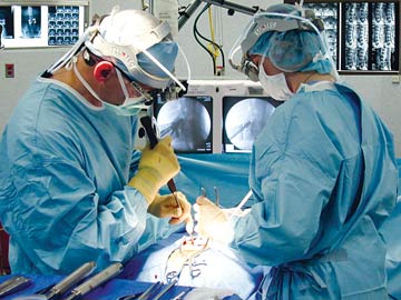
2. Perform separate time outs
BACK TO BASICS Everyone involved in a case should actively participate in processes put in plance to ensure the surgeon operates at the correct spine level.
The main culprit for wrong-site surgeries is a simple counting error made by the surgeon.
To help prevent these mistakes from occurring, conduct three separate time outs during the three localization attempts — pre-incision, post-exposure and at the procedure's completion — to ensure the correct vertebrae has been located and treated. Every member of the surgical team should be engaged in ensuring the correct site is identified. They should stop what they're doing when it's announced a localization attempt is about to start and focus all of their attention on the patient, the surgeon and the intraoperative images that are captured.
After the images are taken, the surgeon counts out loud to the targeted vertebrae level. Everyone in the room should feel empowered to question the surgeon if they don't agree with the count. The decision to begin the operation at the identified site will ultimately be the surgeon's call, but all members of the surgical team shouldn't hesitate to ask for another image and count if there's any doubt about the location of the correct surgical site.
To make this confirmation process work, every person in the OR should receive advanced training on how the localization attempts will be done. They should be familiar with what spine anatomy looks like while viewing live fluoroscopy images on the C-arm monitor. They should know what the C2 and sacrum look like, where they're located and understand their importance in ensuring counts to the correct spine level are correct.
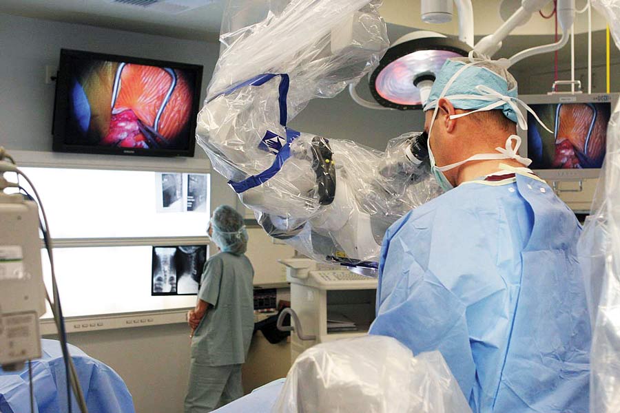
3. Capture the best possible image
CLEAR VIEW Surgeons should locate the correct surgical site based on quality images that leave no doubt about the location of the targeted vertebrae.
The biggest factor that contributes to surgeons counting to the wrong vertebrae is the poor quality of intraoperative images. Some areas of the spine, particularly the thoracic spine, are difficult to visualize and access. It's hard to be confident you're in the right spot, even during the perfect case. The reasons for this could be related to the patient's body type or specific anatomy. The patient could be obese or have abnormal pathology that makes it difficult to properly view the vertebrae. If they have osteoporosis, for example, bones appear very light on X-rays and are therefore hard to see on intraoperative images taken in the OR. Lack of adequate visualization is sometimes a technical issue; the C-arm is simply not producing images that are clear enough to discern individual discs.
Surgeons can't change a person's body habitus or the technical quality of the C-arm they're using in the OR. They can, however, plan in advance of scheduled procedures to ensure they're working with the best possible image in the OR. If a patient is obese or has pathology that might create problems in getting a usable image, surgeons could request to use the best performing C-arm in their facility. They could also consider having a localized marker placed before surgery at the bone they're going to treat.
This pre-operative localization is very helpful for procedures that are in particularly challenging anatomic areas. For example, my patients who are scheduled for thoracic spine procedures visit a radiologist to get a full-body X-ray or CAT scan. During the imaging session, the radiologist places a small coil into the disc that needs repair or removal, so I don't have to count to the correct spot in the OR. I simply capture an intraoperative image to locate the coil and therefore the intended surgical site.
The biggest factor that contributes to surgeons counting to the wrong vertebrae is the poor quality of intraoperative images.
4. Invest in surgical navigation
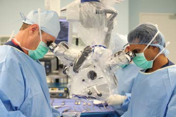
The superior image quality provided by surgical navigation technology can further reduce the likelihood of wrong-site errors occurring. The platforms are expensive and there's a learning curve to operating them proficiently, but the 3D views help to ensure your team is working on the right disc. These imaging systems allow for another level of safety that eliminates the scenario of not being able to capture high-quality intraoperative images, even when operating on a patient with a suboptimal body habitus.
I use 3D navigation for challenging posterior cervical spine surgeries, particularly those performed at the cervical-thoracic junction. Using a three-dimensional CT scan instead of an X-ray in the OR doesn't change the need to localize the surgery site and count to confirm you've located it correctly. However, the images do improve my visualization during all steps of the procedures, and I haven't had a single wrong-site event thanks to my ability to properly identify the correct spine level during these challenging cases.
Focus on the fundamentals
Although advanced technologies can be invaluable in helping surgical teams prevent wrong-site surgery during spine cases, the most reproduceable solution is the localization time outs. There's no big cost associated with performing several time outs during a procedure to confirm the correct site. All it takes is a culture change. Anybody can make a mistake, but it's hard to imagine multiple members of the surgical team making the exact same counting error if there's a culture in place that empowers everyone in the OR to speak up to ensure the surgeon is zeroing in on the right site. OSM
.svg?sfvrsn=56b2f850_5)