January 7, 2021

Share:
eNews Briefs January 7, 2021

THIS WEEK'S ARTICLES

Swiss Hospital Performs World's First Holographically-Navigated Spine Surgery

New Spinal Fusion Approach 'Flips' the Script

Coordinated Care Pathway Pays Off

Determining the True Nature of Bone Density

Multimodal Pain Management Works Well for Spine


Swiss Hospital Performs World's First Holographically-Navigated Spine Surgery

Another landmark in the advancing field of surgical augmented reality (AR).
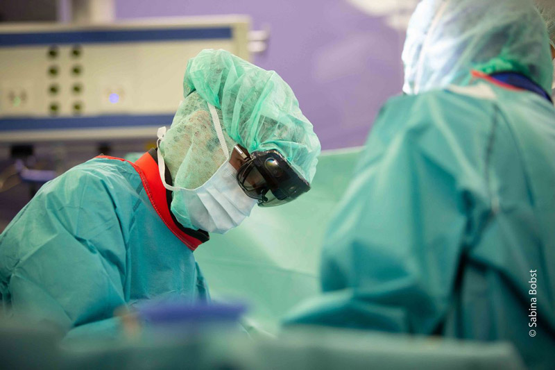
GOGGLES ON Credit: Sabina Bobst/Balgrist University Hospital
GOGGLES ON The surgeon uses augmented reality technology to overlay 3D holographic images on top of real patient anatomy.

Augmented reality (AR) in the OR has taken a significant step forward with the news that a team at Balgrist University Hospital in Zurich, Switzerland, successfully completed the world's first holographically-navigated spine surgery. Performed in early December, the procedure was part of a randomized clinical study based on technology developed at the hospital in conjunction with Microsoft.

The surgical team was led by Mazda Farshad, MD, MPH, chief of orthopedics and spine surgery at Balgrist. The patient suffered from lower lumbar spine degeneration, a significantly narrowed spinal canal, and the strong pain and sensory disorders in the legs associated with that condition.

Dr. Farshad wore goggles powered by AR navigation software to view 3D representations of the patient's affected anatomy generated from preoperative CT images. The images were projected onto the surgical field, overlaying the patient's real anatomy. For example, the exact insertion point and trajectory of a screw was shown directly on the patient's anatomy. Dr. Farshad reported that the holographic images enhanced his senses and improved his perception. According to Microsoft, the patient is now symptom-free and doing well.

Philipp Fürnstahl, MS, PhD, head of the Research in Orthopedic Computer Science group at Balgrist, described the surgery as "an eminent milestone toward orthopedics shaped by computer technology with the goal of fully digitized treatment." Watch a video about the surgery here.

New Spinal Fusion Approach 'Flips' the Script

Single-position anterior-posterior lumbar fusion surgery (SPLS) removes the need to reposition the patient midway through the procedure.

EASY ACCESS Credit: Juliana Thomas/NYU Langone Health
EASY ACCESS Study co-author Themistocles Protopsaltis, MD, chief of NYU Langone's division of spine surgery, believes SPLS results in quicker operations and faster patient recoveries.

Researchers at NYU Langone Health have developed a new spinal fusion surgery technique that claims to offer improved outcomes, reduced OR time and shorter lengths of stay for patients. The novel approach, dubbed "single-position anterior-posterior lumbar fusion surgery" (SPLS), is also reported to reduce perioperative blood loss and postoperative ileus (intestinal blockage), as well as intraoperative radiation dosage.

Spinal fusion has traditionally been performed by repositioning or "flipping" the patient midway through surgery from their back or side to their stomach. SPLS, conversely, allows the entire operation to be completed with the patient laying on their side. NYU Langone says this enables the surgical team to approach the spine through both the front and back, leading to a higher fusion rate while minimizing the length of surgery and facilitating a more rapid recovery.

Aaron J. Buckland, MD, lead author of the study and an associate professor in NYU Langone's department of orthopedic surgery, calls SPLS a "practice-changing technique" that's just as safe and effective as the traditional method, with added benefits. "With the improved operative efficiencies of single-position surgery, we are performing less invasive procedures, producing better outcomes and improving safety," he says.

The SPLS study group reviewed nearly 400 perioperative and 90-day outcomes of patients undergoing spinal fusion between May 2012 and June 2019. Of the 397 surgeries, 153 were classified as "flip" procedures and 244 as SPLS procedures. Surgeons at NYU Langone's Spine Center found SPLS reduced operating times from an average of 306 minutes to 103 minutes. Length of hospital stay for the SPLS patients was reduced from an average of four days to two. Reductions in ileus were attributed to reduced time under anesthesia, more minimally invasive abdominal incisions, reduced pain medication requirements, more rapid mobilization and the fact that SPLS patients don't lay face down.

With their retrospective review complete and published in The Spine Journal, the research team is now opening a prospective multicenter study to evaluate SLPS against minimally invasive posterior fusion in order to further validate its efficacy.

Coordinated Care Pathway Pays Off

Early intervention for vertebral compression fractures results in cost savings for hospitals and payors.

Brian Thomas Credit: Brian Thomas
Brian Thomas, vice president of Ortho, Neuro & Spine at Swedish Medical Center, is instituting a coordinated care pathway for patients with painful vertebral compression fractures at Swedish Medical Center in Denver, Colo.

Instituting a coordinated care pathway for patients with painful vertebral compression fractures (VCFs) is paying off at Swedish Medical Center in Denver.

"We have reduced the probability of VCF patients staying overnight at the hospital or being transferred to a skilled nursing facility for multiple nights," says Brian Thomas, vice president of Ortho, Neuro & Spine at Swedish Medical Center. "We also have fewer patients returning to the emergency department two or three times in intractable pain. Our new approach based on early intervention is giving these patients a solution to their pain, and it's resulting in a cost savings for the hospital and for the payors."

That new approach uses a treatment algorithm for VCFs based on recommendations published in 2018 by a multispecialty panel of experts using the RAND Appropriateness Method.1 The VCF Care Pathway adopted by Swedish Medical Center streamlines the process for treating patients with vertebral augmentation via KyphonTM Balloon Kyphoplasty.

Balloon Kyphoplasty is a minimally invasive surgical procedure for the treatment of pathologic vertebral compression fractures due to osteoporosis, cancer or noncancerous tumors.

"At Swedish, we are so convinced of the benefits of kyphoplasty that we have a team of people to coordinate patient care," Thomas says. "First, it can provide immediate pain relief with very little if any prescribed pain medication. Second, it also limits further spine complications and limits further spine complications and improves function.2 This is important because patients with untreated fractures face a downward spiral of health problems and an increased risk of mortality.3-5 And third, although the patient is not guaranteed that they won't get another compression fracture, the odds are in their favor that it will be significantly delayed."

Thomas recommends the following steps for a hospital to institute a VCF Care Pathway:

Engage industry: "I've found that my vendor partners have an enormous amount of experience that has helped us more quickly identify issues and brainstorm potential solutions," he says.

Foster collaboration: "Educate your healthcare community on the dangers of leaving VCF untreated. Share research with physical therapists, assisted living facilities, primary care physicians and home care providers."

Drive down costs: "Engage Accountable Care Organizations (ACO's) if they are prevalent in your community. Show them the differences in the cost of care for a return visit to the emergency department and long-term care vs. kyphoplasty."

To learn more about VCF Care pathway, visit medtronic.com/vcfcarepath.

Note: The following testimonial contains the opinions of Brian Thomas. The opinions and practice presented herein are for information purposes only and the decision of which techniques to use in a particular surgical application should be made by the surgeon based on the individual facts and circumstances of the patient and previous surgical experience.

References

1. Hirsch JA, Beall DP, Chambers MR, et al. Management of vertebral fragility fractures: a clinical care pathway developed by a multispecialty panel using the RAND/UCLA Appropriateness Method. Spine J. 2018. doi: 10.1016/j.spinee.2018.07.025.

2. Boonen, S. et al, Journal of Bone and Mineral Research, 2011 July (Vol. 26, No. 7): 16271634.

3. Schlaich C et al. Reduced pulmonary function in patients with spinal osteoporotic fractures. Osteoporos Int. 1998;8(3):261-7.

4. Gold DT. The clinical impact of vertebral fractures: quality of life in women with osteoporosis. Bone. 1996;18(3 Suppl):185S-189S.

5. Old J, Calvert M. Vertebral Compression Fractures in the Elderly. Am Fam Physician. 2004 Jan 1;69(1):111-6.

Determining the True Nature of Bone Density

Hospital for Special Surgery team investigates 3D tool to better visualize bone abnormalities before spinal fusion surgeries.

A new bone study of 54 patients has identified a link between skeletal abnormalities and the likelihood of complications after spinal fusion procedures. This is no small finding, as researchers estimated that approximately 45% of the 400,000 U.S. patients who undergo these procedures each year experience postoperative complications that are often directly related to the bone's inability to tolerate the hardware surgeons use to support the skeleton.

Researchers at the Hospital for Special Surgery (HSS) in New York City identified issues with dual X-ray absorptiometry (DXA) technology, which many surgeons use to evaluate patients' bones before spine fusion surgery is called. They say DXA imaging only provides a "rough sense" of bone strength, and that patients whose DXA scans appear normal often turn out to actually have bones so weak that the appliances used during spinal fusion are at risk of failure. "On DXA, the spine is almost always going to look fine, which can be misleading," says HSS endocrinologist and bone specialist Emily Stein, MD, whose team set about looking for a more sensitive measure of bone quality that can better identify abnormalities.

They believe they've found that more sensitive tool in a "cutting-edge" technique called high-resolution peripheral quantitative computed tomography (HR-pQCT). This technique can separately measure how much bone is in the outer (cortical) and inner (trabecular) compartments, then measure microscopically how the trabecular bone network is organized, including the number, thickness and spacing between the parts of the trabeculae network. This "microarchitecture" approach, they say, may provide a more accurate assessment of skeletal health than DXA, particularly with patients "who frequently have changes in the area of the fusion which undermine the utility of DXA." Essentially, DXA provides a 2D measure of bone density, while HR-pQCT provides a 3D measure.

Of the 54 patients in the study, 14 had experienced complications within the first six months of surgery, including broken rods, loosened bone screws, fractures and abnormal bending of the spine. The researchers discovered that those with abnormalities found on HR-pQCT, but not on DXA, were significantly more likely to experience complications involving lower bone mineral density in the trabeculae, fewer and thinner trabeculae and thinner cortices.

Unfortunately, HR-pQCT is a research tool not yet widely available to surgeons, but it's pointing the HSS researchers toward a better understanding of individual patients' spinal anatomy. Dr. Stein's group is expanding its study. "We want to have the most optimal strategies for lowering complications in our patients, and that begins with understanding who is at risk and why," she says. "In our work with additional patients, we hope to define which features, or group of features, of the bone structure is most important in contributing to surgical success. This will help us to devise the most targeted treatment strategies for our patients."

The team published its initial findings last month in Bone.

Multimodal Pain Management Works Well for Spine

Positive patient outcomes persisted after Michigan's tough opioid-prescribing laws went into effect.

A data analysis of the impact Michigan's restrictive 2017 opioid-prescribing laws has had on spine surgery patients has uncovered information about changes in preoperative narcotic use, short-term outcomes and readmission rates after spinal surgery. All signs point to the effectiveness of opioid-sparing pain management techniques.

Patient data from a year before and a year after initiation of the laws was queried from the Michigan Spine Surgery Improvement Collaborative database. The approximately 12,000 patients in each group generally had similar demographic and surgical characteristics.

The analysis found that the percentage of patients taking daily narcotics preoperatively before and after the laws went into effect decreased from 48.7% to 39.7%. However, no significant differences were identified between the groups three months after surgery in terms of minimum clinically important difference, numeric rating scale (NRS) score of back and leg pain, patient satisfaction or 90-day readmission rate.

"There was a meaningful decrease in preoperative narcotic use, but notably there was no apparent negative impact on postoperative recovery, patient satisfaction or short-term outcomes after spinal surgery despite more restrictive opioid prescribing," say the authors, whose study was posted online in the Journal of Neurosurgery ahead of print.

The findings fall in line with a growing body of anecdotal and data-driven evidence that suggests multimodal pain management techniques can serve as an effective alternative to opioids.

Related Articles