January 21, 2021

Share:
eNews Briefs January 21, 2021

THIS WEEK'S ARTICLES

Five Tips for Reducing Delirium Risk in Older Patients

Biofilm on Retrieved Implants Prevalent Among Pseudarthrosis Patients

Early Intervention is Key to Successful Treatment of Painful Vertebral Fractures

Bone Mineral Density Measurements Often Miss Fracture Risks

3D Printing Improves Spine Surgery Outcomes


Five Tips for Reducing Delirium Risk in Older Patients

The new recommendations have minimal impact on your bottom line and positively impact patient outcomes.

TEAM EFFORT 
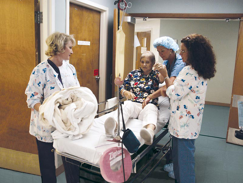
TEAM EFFORT All perioperative staff have a role to play in helping to reorient patients after surgery to reduce the risk of delirium.

Older individuals are at heightened risk of developing debilitating post-op delirium or neurocognitive disorder, which can lead to delayed discharge, increased complications and higher healthcare costs. To prevent cognitive impairment in geriatric patients, follow these risk-reduction recommendations issued by the American Society of Anesthesiologists' (ASA) Perioperative Brain Health Initiative (PBHI).

In its new consensus review, published in the British Journal of Anaesthesia, PBHI identifies five critical steps that should be taken throughout the perioperative process to reduce the risk of delirium in older patients:

  • Increase awareness. Anesthesiologists should guide training and education initiatives for all staff who work with older patients to better identify and manage patients with delirium.
  • Screen preoperatively and postoperatively. Perform preoperative baseline cognitive screening using a recognized test on all older adult patients before surgery as well as before they leave the recovery unit.
  • Non-pharmacologic interventions. The entire perioperative care team should work together to get patients walking, periodically orient them to where they are and provide physical therapy. Return glasses, dentures and hearing aids to patients as soon as possible after surgery.
  • Pain control. Anesthesiologists and surgeons should work together to optimize postoperative pain control, preferably using minimally sedating options and multimodal techniques.
  • Avoidance of antipsychotic drugs. Don't treat delirium with antipsychotic drugs and benzodiazepines as a first response. Instead, enlist family members to help reorient the patient and focus on pain relief and other treatable factors. All staff should understand and follow this approach.

None of BPHI's recommendations require purchases of new equipment or new drugs, notes Carol J. Peden, MD, lead author of the recommendations and an adjunct professor of clinical anesthesiology at Keck Medicine of the University of Southern California and the University of Pennsylvania. To minimize delirium risk, she emphasizes that anesthesiologists must lead the process and the entire perioperative staff must buy in.

"Reducing the incidence of delirium is not in the hands of anesthesiologists alone, but we are well-placed to help lead the organizational initiatives needed to address the problem," she says.

Read the full recommendations here.

Biofilm on Retrieved Implants Prevalent Among Pseudarthrosis Patients

Loose screws could be due to infection rather than mechanical failure.

NOT SO ASEPTIC Credit: Aakash Agarwal, PhD
NOT SO ASEPTIC Researchers found biofilm on retrieved implants from aseptic revision surgeries.

Recent studies have associated pseudarthrosis among spine surgery patients and its attendant pedicle screw loosening with subchronic infection at the pedicle of the vertebra, which in turn often leads to bacterial contamination. Unfortunately, most of these infections from biofilms on "aseptic" screw loosening go undiagnosed, as they present no known clinical symptoms. Many surgeons simply view the problem as a mechanical failure at the screw-bone interface.

A team of researchers from the U.S. and India undertook a first-of-its-kind study to visually capture the architecture of these undiagnosed infections by examining explants from patients undergoing revision spine surgery for pseudarthrosis. The explants were fixed in glutaraldehyde solution and then imaged using scanning electron microscopy and X-ray spectroscopy to evaluate the biofilm architecture. Eight patient swabs from tissues around the implants were sent for cultures to assess bacterial infiltration in tissues beyond the biofilm.

According to the study, 77% of pseudarthrosis cases had loose pedicle screws and 72% showed biofilm on the implants. In addition, areas with biofilms consistently tested negative for calcium phosphate, which aids in bone mineralization, while those without biofilms tested positive for the mineral. Intraoperative diagnosis of implant infections is complicated by the fact that tissue and swab cultures of the surrounding screws do not efficiently demonstrate bacteria growth.

The researchers, referring to "the biological pathway hypothesis," concluded, "In the absence of the clinical presentation of infection, impregnated bacteria could form a biofilm around an implant, and this biofilm can remain undetected via contemporary diagnostic methods, including swabbing. Implant biofilm is frequently present in 'aseptic' pseudarthrosis cases." They hope the study illuminates "the surreptitious dynamics of implant and impregnated microbes, and possibly explains the sudden onset of delayed and late infection responses," which can occur from 90 days to many years after surgery.

They added that the findings highlight the importance of keeping screw/screw-bone interfaces devoid of bioload because of the propensity of bacterial inoculation to form biofilms around the implant.

"This discovery concludes that the risk of bacterial contamination during spine surgery cannot alone be measured by the rates of acute infection, and therefore preventative measures to avoid all possibly known modes of contamination of the surgical site and the implantable device is crucial for the long-term safety of the patient, spanning many years," said study author Aakash Agarwal, PhD.

Dr. Agarwal, an adjunct faculty member of the departments of bioengineering and orthopedic surgery at the University of Toledo (Ohio)'s Colleges of Engineering and Medicine, says he requested that the FDA mandate spinal implants be sold as individual, "truly single-use and pre-sterile devices" with custom, implant-specific intraoperative guards to avoid exposure until implantation. He based that request on findings from a Level II clinical study he led that was published in October.

Early Intervention is Key to Successful Treatment of Painful Vertebral Fractures

Minimally invasive surgery offers a solution for patient pain and discomfort.

Sarat Piduru, MD Credit: Dr. Sarat Piduri, MD.
CONSISTENT APPROACH. Sarat Piduru, MD, championed a new care pathway utilizing Kyphon Balloon Kyphoplasty to treat VCF patients.

Vertebral compression fractures (VCF) are the most common type of fragility fracture, occurring more than 800,000 times a year in the United States alone.1 VCFs often cause debilitating back pain leading to reduced function. Diagnosis and treatment of VCFs is challenging because many providers who interact with VCF patients are not familiar or experienced with treatment options beyond conservative management, which includes bed rest, pain medication and use of a back brace.

In order to improve care of patients with painful VCFs, a multispecialty panel of experts published a VCF care pathway in 2019. The care pathway seeks to streamline patient access to early intervention via vertebral augmentation. This can reduce the cycle of pain, limit the downward spiral of complications, and lower mortality risks.

Dr. Sarat Piduru is an interventional radiologist who championed the care pathway at Eastside Medical Center in Snellville, Ga.

"Having the care pathway means there is a consistent approach to treating patients who come to the hospital with painful vertebral compression fractures," Piduru says. "With everyone on the same page, we can send patients home more quickly after they present at the emergency room, knowing that there is a plan in place to get them scheduled for vertebral augmentation. This helps us avoid keeping patients in the hospital for days on pain control and then having them readmitted days later because they weren't treated for the underlying cause of the pain."

Piduru uses Kyphon Balloon Kyphoplasty to provide patients with an immediate solute to reduce pain and discomfort caused by their VCF.2 Kyphon Balloon Kyphoplasty is a minimally invasive surgical procedure for the treatment of pathological fractures of the vertebral body due to osteoporosis, cancer or noncancerous tumors.

"An important part of implementing the VCF care pathway has been educating the referring physicians as well as nurses," Piduru says. "We tell them we would like to treat patients as soon as possible after they suffer a VCF. There's no sense in making patients wait in pain when there is an effective treatment available that can bring rapid pain relief like balloon kyphoplasty."

This solution prevents a host of downstream effects of leaving a VCF untreated, from digestive issues to blood clots to pneumonia.3

"These patients with vertebral compression fractures are mostly older people, and the pain can really limit their activities, not just exercise and hobbies but even basic things like walking to the bathroom or getting dressed," Piduru says. "Many of them spend days or weeks in bed or in an easy chair because of the pain. Treating the patients to relieve their pain helps them get moving again and can avoid this downward spiral of complications."

*The following testimonial contains the opinions of and personal surgical techniques practiced by Sarat Piduru, MD. The opinions and techniques presented herein are for information purposes only and the decision of which techniques to use in a particular surgical application should be made by the surgeon based on the individual facts and circumstances of the patient and previous surgical experience.

References

1. Medtronic data on file.

2. Boonen, S. et al, Journal of Bone and Mineral Research, 2011 July (Vol. 26, No. 7): 1627-1634.

3. Hirsch JA, Beall DP, Chambers MR, et al. Management of vertebral fragility fractures: a clinical care pathway developed by a multispecialty panel using the RAND/UCLA Appropriateness Method. Spine J. 2018. doi: 10.1016/j.spinee.2018.07.025.

Bone Mineral Density Measurements Often Miss Fracture Risks

Supplemental use of other screening tools could better identify the need for prompt intervention in postmenopausal women with osteoporosis.

One-third of recently postmenopausal women who are diagnosed with osteoporosis are at high risk for fracture, according to a new study. The findings suggest prompt interventions, including kyphoplasty and vertebroplasty, may be the best treatment option for these patients.

The team of Canadian and Iranian researchers examined whether balance and functional mobility independently predict bone mineral density (BMD) in postmenopausal women. They measured BMD at the hip and spine with dual-energy X-ray absorptiometry (DEXA) in three 20-patient groups: those with osteoporosis; those with osteopenia and thus at high risk for osteoporosis; and those with normal BMD. All patients performed the single leg stance (SLS), timed-up-and-go (TUG) and six-meter walking tests. The researchers found that three factors predicted low BMD: less time to hold the SLS, longer TUG time and older age.

"The SLS and TUG independently predicted BMD among women undergoing recent menopause with osteoporosis, osteopenia and normal BMD," the team reported in the journal Geriatric Nursing. "Our research suggests that incorporating the SLS and TUG into risk assessments for postmenopausal women may facilitate prompt and targeted intervention."

The researchers cited literature that notes approximately one-third of postmenopausal women develop osteoporosis, leading to annual direct medical costs of nearly $20 billion. Relying on BMD measurements alone to diagnose the condition is a risky venture, they say, as error rates can occur as frequently as one in five patients. "Impaired balance and functional mobility are modifiable risk factors that are prevalent among postmenopausal women with osteoporosis," the team writes. "Therefore, they may serve as viable screening measures for postmenopausal women with osteoporosis and osteopenia."

While the researchers say more research on a larger and more diverse patient population is required, their work provides more evidence that effective screening and early intervention can help providers better treat postmenopausal women afflicted with the condition.

3D Printing Improves Spine Surgery Outcomes

Creating precise models of patient anatomy can simplify complex procedures.

The use of 3D imaging software to plan and execute spine surgeries continues to gain favor, but what about using 3D-printed models of spine anatomy to help simulate and plan a procedure? That's happening, too, and was a topic of discussion during a session at the recent annual North American Spine Society (NASS) meeting.

One panelist from that session is intrigued by the potential for 3D printing to produce better, more accurate outcomes. "I have some interest in using it for my patients," says Srinivas K. Prasad, MD, MS, an associate professor of neurological and orthopedic surgery at Thomas Jefferson University in Philadelphia, who notes that 3D printers are commercially available for $5,000 or $6,000. The materials used by the printer to create the model are an additional cost, but Dr. Prasad says it's minimal. "You can start printing models of patient anatomy for [around] a few hundred dollars," he says.

Dr. Prasad notes that because this application is a fairly recent development, spine experts are working on developing protocols, such as exactly how patient data sets should be plugged into the printer, as well as the right materials to use with the printer to get more accurate models of the spine.

While printing a 3D model of a patient's spine might seem like overkill for most cases, especially considering the availability of 3D modeling and simulation software, it can be extremely useful in critical or complex cases. Norton Children's Hospital in Louisville, Ky., last year revealed that one of its surgeons used 3D printing to treat a 13-year-old Indiana girl with achondroplasia, a common form of dwarfism, and a spinal deformity.

Orthopedic surgeon Kent L. Walker, DO, eyeing the complexity and potential danger of the intervention, converted CT images of the girl's spine into digital files that he fed into a 3D printer. Out came an exact physical replica of her spine, and he even created custom-made drill guides for each of the affected vertebra. "The result of the 3D-printed models and guides is increased accuracy and increased safety putting screws in," says Dr. Walker. "Having the ability to practice before the procedure makes the surgery safer and more effective."

This video shows how St. Louis Children's Hospital worked with Washington University's Medical 3D Printing Center to produce a model of the spine of a young patient with spina bifida. While this case, along with the previously described case, were inpatient procedures, it's easy to see that 3D printing could prove valuable to outpatient centers going forward as ever-more-complex cases migrate from hospitals.

Related Articles