Performing Cataract Surgery in Comfort

Share:

Heads-up visualization systems are ergonomic solutions for surgeons that have the potential to improve visual outcomes.


Ophthalmology is a physically demanding specialty. Surgeons spend good portions of their day looking through the eyepieces of surgical microscopes, leaning forward in unnatural positions while performing delicate microsurgery that requires minute hand movements. They adjust their chairs and reposition the patient, but are often forced to endure ergonomically unfavorable positions that put stress on their back, neck, arms and even their eyes.

I’ve been practicing for 20 years and perform between 75 and 100 cataract surgeries a week. Although each procedure takes me only about five minutes to complete, the cumulative effects of ergonomic stress can take their toll. The repetitive nature of working in ophthalmology has been shown to cause long-lasting degenerative and physiologic changes to the vertebrae that lead to scoliosis, bulging discs and arthritis of the neck and spine in the lumbar thoracic and cervical regions. Many surgeons have suffered short- or long-term disabilities throughout their careers.

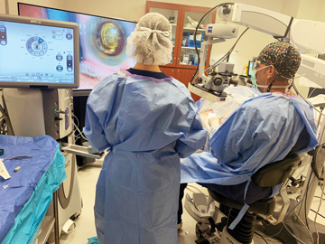
These concerns led me to adopt heads-up 3D visualization, which frees me from sitting behind a surgical microscope for hours at a time. I’ve had lower back issues for most of my adult life and underwent a microdiscectomy due to sports-related injuries. It’s important for me to be in a comfortable position when I operate to protect my back and guard against developing other chronic issues. I’ve experienced significantly less neck and back pain since performing heads-up cataract surgery, but that’s only one of the reasons I rely on the technology at my practice.

Plenty of pluses

It’s very difficult for ophthalmic surgeons to be disciplined enough during a day packed with fast-moving cataract cases to put their body in the right ergonomic position every time. This is where heads-up 3D visualization systems help tremendously. Surgeons don’t need to lean forward or stretch their neck awkwardly to reach the microscope’s oculars; they don a special headset that lets them see 3D images of surgery projected onto a large flat screen monitor. They can sit back, shift their weight and move their head and neck, putting themselves in more comfortable positions instead of sitting in a static position as they operate, which over time can cause back and neck muscles to spasm.

Working with a heads-up display is easier on the eyes than looking through a surgical microscope and 3D imaging technology offers a greater depth of field. It also allows for more collaboration among members of the surgical team, who can see exactly what’s going on during the case and prepare for the next steps. In that regard, heads-up displays can help make surgery more efficient and more fluid.

I’ve found that the aches and pains I feel while sitting behind a microscope can be distracting when I’m performing the small movements of cataract surgery. Although it hasn’t yet been quantified, I truly believe that I’m more likely to make a mistake while operating in pain, perhaps because I’m subconsciously rushing through steps in the surgery to get relief from the soreness. Surgeons who operate using heads-up 3D displays are more comfortable as they’re working, and I believe that translates to better outcomes.

Heads-up technology is also easier to set up between cases. Instead of needing to make small adjustments to the surgical microscope, seat and bed to ensure comfortable access to and visualization of the patient’s eye, surgeons can walk into the OR, sit down and start operating. You end up saving about a minute or two between cases. In high-volume facilities, these precious few minutes can quickly add up.

Does Less Soreness Lead to Safer Surgery?
RESEARCH RESULTS
BIG SCREEN BENEFITS Performing surgery in 3D lets surgeons focus on the task at hand instead of their physical discomfort.

My recent study published in the Journal of Cataract and Refractive Surgery compared the outcomes of 2,320 cataract procedures performed with traditional surgical microscopes or 3D heads-up displays (osmag.net/cataracts). Patients in both groups underwent surgery with femtosecond laser assistance or traditional phacoemulsification. Patients who underwent 3D surgery suffered 12 complications (0.72%), compared with five complications (0.77%) among patients in the microscope group. The mean surgical time in each group was 6.48 and 6.52 minutes, respectively.

Heads-up visualization seems to offer similar safety and efficiency as the traditional binocular microscope, according to the study’s findings. Still, I believe the use of heads-up displays will eventually lead to safer surgery because surgeons who operate in ergonomic comfort are better able to focus on the intricacies of performing delicate eye procedures.            
Robert Weinstock, MD

Seeing a difference

TEAM EFFORT Heads-up visualization allows for more collaboration during surgery because other people in the operating room can track the progress of the case.

I’m often asked to justify the cost of investing in heads-up technology — platforms cost between $75,000 and $250,000, depending on the features — because you can’t bill a patient or Medicare for its use. My response is you can’t put a price tag on your health. Surgeons who lose a month or two of work due to repetitive strain injuries cost their practice and surgery center hundreds of thousands of dollars in revenue. Surgeons who are forced to end their careers several years earlier than expected because of chronic neck and back issues could lose millions of dollars in personal income. Even surgeons who operate through the pain aren’t physically able to work as efficiently or productively. I think it’s best to invest in equipment that helps surgeons achieve clinical success over the long term. Heads-up 3D visualization falls into that category.

The technology is innovative, so some surgeons who are accustomed to operating with a surgical microscope might push back against using it. However, I’ve found that progressive surgeons are perfectly capable of adapting to performing heads-up surgery. 

Surprisingly, there has never been a lot of focus placed on proper ergonomics during schooling or training for a career in ophthalmology. Some experienced physicians teach residents and young surgeons how to adjust and tilt the microscope in different positions and be conscientious about their posture when they perform surgery. Still, more young surgeons should take surgical ergonomics seriously and understand the risks involved in poor body positioning, including the potential shortening of their careers. I think it’s important for them to give 3D visualization a try, because it’s often difficult to convince experienced physicians to alter how they perform surgery. I know firsthand that once a surgeon notices the difference in how their body feels after heads-up surgery, it’s easy to convince them to make the switch. OSM

Related Articles

Wired for Success

In her 24 years as a nurse at Penn Medicine, Connie Croce has seen the evolution from open to laparoscopic to robotic surgery....

To Optimize OR Design, Put People First

Through my decades of researching, testing and helping implement healthcare design solutions, I’ve learned an important lesson: A human-centered and evidence-based...