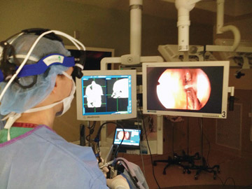
The latest navigation systems are driven by electromagnetic guidance (with sensors attached to or embedded in the instruments) where the system is linked from an image processor through direct connection to the surgical instruments being used.
It's this direct link that allows surgeons to navigate seamlessly through the nasal cavity without maintaining a direct line of sight between the instruments and the image processor. One caveat: These systems require surgeons to use proprietary
instruments —something that could affect surgeons who are partial to the instrumentation they've always used.
Surgeons need image-guided navigation for complex procedures involving significant nasal and sinus pathology, or distorted or aberrant anatomy.
Although most of today's image-guided systems employ electromagnetic guidance, older systems still use infrared guidance efficiently. This infrared guidance comes in two forms:
- Passive. Fiducial markers are placed on the patient and use instruments that reflect infrared light back to the system's camera.
- Active. Infrared-emitting diodes are placed on surgical instruments that are tracked with an overhanging camera.
While any type of image-guided technology is better than none at all, I'm partial to electromagnetic guidance ?—largely because of its accuracy and because, unlike infrared guidance, it doesn't require me to maintain a clear line of sight
between the instruments and the imaging unit. The process begins with patients receiving pre-op CT scans, which are loaded into the system's 3D navigator. Once the images are loaded, we calibrate the patient's actual facial structure to the
radiologic images, and the computer matches the two up through an algorithm. This process allows us to map out the optimal route to the anatomy we need to access, and the electromagnetic tracking allows us to guide our instruments safely and
efficiently along the preplanned surgical pathway.
Obviously, image guidance doesn't replace the superior skill and technique of surgeons who perform delicate ENT procedures, nor does it completely eliminate the risk of complications. However, the technology is an effective tool that lets surgeons
identify and avoid areas of the nasal cavity that, if breached, could cause disastrous outcomes: the sphenoid sinus, which is in close proximity to the carotid artery and optic nerve; the roof of the ethmoid, an area separated from the brain
by a very thin plate of bone; and the lamina papyracea, a structure that separates orbits from the sinuses.
These fragile areas can be anatomically distorted from previous surgeries or impacted by disease. Without the additional visualization provided by image-guided technology, even the most experienced otolaryngologists would hesitate to get too close.
The real-time visualization of image guidance lets surgeons successfully treat pathology that they otherwise wouldn't be able to —and the true beneficiary of this technology is the patient.
Image guidance has been a game-changer for patients with severe nasal polyposis (non-cancerous growths lining the nasal passages) and severe chronic rhinosinusitis (a painful inflammation of the paranasal sinuses and linings of the nasal passages
that lasts for 12 weeks or longer). It's also helped tremendously in treating patients with sinonasal tumors. Surgeons need image-guided navigation for complex procedures involving significant nasal and sinus pathology, or distorted or aberrant
anatomy.
In addition to improved precision and ultimately better surgical outcomes, image-guided systems allow surgeons to operate more efficiently, which reduces surgical times and ensures patients spend less time under anesthesia.
.svg?sfvrsn=be606e78_3)
.svg?sfvrsn=56b2f850_5)